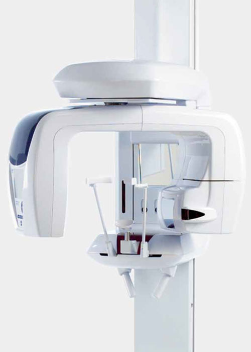
J Morita Veraviewepocs 3D F80
- Exceptional image clarity
- Multifunctional: 3D, panoramic & cephalometric
- Enhanced patient safety with Dose Reduction Feature
- Three options for easy & accurate 3D positioning
- Seven pre-programmed panoramic functions with magnification options
- Fast scan times: 7.4 second panoramic; 9.4 second 3D scan
- FOV: Ø 40 x H 40 mm, Ø 40 x H 80 mm, Ø 80 x H 50 mm, Ø 80 x H 80 mm
Fields of View
Our imaging equipment offers the option of 4 possible exposure sizes, ranging from Ø 40 x H 40 mm up to Ø 80 x H 80 mm, allowing reduced radiation dose yet exceptional clarity of the images.
This model’s small and medium fields of view are well suited for endodontics, periodontics, and general dentistry.
Easy 3D Positioning
Flexibility: Veraviewepocs offers flexibility in positioning methods. The
region of interest can be positioned by the panoramic image, the bi-directional scout, or the 5 positioning laser beams.
Panoramic Image with Scout Feature: Before taking a 3D image, a high-resolution panoramic exposure is taken to target the region of interest on the PC monitor. The C-arm will automatically move into the
optimum patient position to get 3D images at the center of the region of interest.
Bi-directional Scout: After initial positioning is accomplished by the 3
positioning laser beams, bidirectional X-ray images can be taken to confirm that the position is accurate. If it is not, simply adjust the position of the image on the computer by placing the cursor at the center of the region of interest.
Direct Positioning with 5 Laser Beams: Five positioning laser beams set the patient‘s position and align the region of interest. First, the patient‘s initial position is set using the 3 laser beams. Then, 2 additional laser beams are aligned to the region of interest. The C-arm will automatically move to the right position.
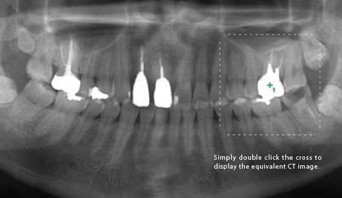
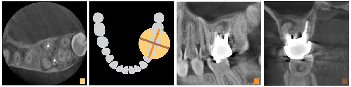
Clinical Case Example: The panoramic image suggests that there is an apical lesion on the distal root of tooth #26. Further inspection with a 3D image, however, shows that the lesion is on the buccal side of an extremely curved mesial root.